Cancer cells are cells that divide relentlessly, forming solid tumors or flooding the blood with abnormal cells. Cell division is a normal process used by the body for growth and repair. A parent cell divides to form two daughter cells, and these daughter cells are used to build new tissue, or to replace cells that have died because of aging or damage. Healthy cells stop dividing when there is no longer a need for more daughter cells, but cancer cells continue to produce copies. They are also able to spread from one part of the body to another in a process known as metastasis.[1]
Major Characteristic: Immortality
Cancer cells have unique features that make them “immortal” according to some researchers. The enzyme telomerase is used to extend the cancer cell’s life span. While the telomeres of most cells shorten after each division, eventually causing the cell to die, telomerase extends the cell’s telomeres. This is a major reason that cancer cells can accumulate over time, creating tumors.
Typology (or Phenotypes) of Cancer Cells
There are different categories of cancer cell, defined according to the cell type from which they originate.[2]
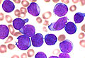
Leukaemia, originate in the tissues responsible for producing new blood cells, most commonly in the bone marrow.
Lymphoma and myeloma, derived from cells of the immune system.
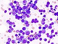
Myeloma, derived from cells of the immune system.

Sarcoma, originating in connective tissue, including fat, muscle and bone. Central nervous system, derived from cells of the body and spinal cord.
Mesothelioma, originating in the mesothelium; the lining of body cavities.

Histological features of normal cells and cancer cells
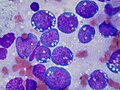
Cancer cells have distinguishing histological features visible under the microscope. The nucleus is often large and irregular, and the cytoplasm may also display abnormalities.[3]
The shape, size, protein composition, and texture of the nucleus are often altered in malignant cells. The nucleus may acquire grooves, folds or indentations, chromatin may aggregate or disperse, and the nucleolus can become enlarged. In normal cells, the nucleus is often round or ellipsoid in shape, but in cancer cells the outline is often irregular. Different combinations of abnormalities are characteristic of different cancer types, to the extent that nuclear appearance can be used as a marker in cancer diagnostics and staging.[4]
-
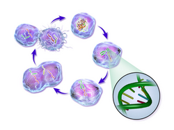
Life cycle of a cancer cell.
Carcinogenesis
Cancer cells are created when the genes responsible for regulating cell division are damaged. Carcinogenesis is caused by mutation and epimutation of the genetic material of normal cells, which upsets the normal balance between proliferation and cell death. This results in uncontrolled cell division and the evolution of those cells by natural selection in the body. The uncontrolled and often rapid proliferation of cells can lead to benign or malignant tumours (cancer). Benign tumors do not spread to other parts of the body or invade other tissues. Malignant tumors can invade other organs, spread to distant locations (metastasis) and become life-threatening.
More than one mutation is necessary for carcinogenesis. In fact, a series of several mutations to certain classes of genes is usually required before a normal cell will transform into a cancer cell.[5]
Damage to DNA can be caused by exposure to radiation, chemicals, and other environmental sources, but mutations also accumulate naturally over time through uncorrected errors in DNA transcription, making age another risk factor. Oncoviruses can cause certain types of cancer, and genetics are also known to play a role.[6]
Stem cell research suggests that excess SP2 protein may turn stem cells into cancer cells.[7] However, a lack of particular co-stimulated molecules that aid in the way antigens react with lymphocytes can impair the natural killer cells’ function, ultimately leading to cancer.[8][not in citation given]
The Immune System
Cells playing roles in the immune system, such as T-cells, are thought to use a dual receptor system when they determine whether or not to kill sick or damaged human cells. If a cell is under stress, turning into tumors, or infected, molecules including MIC-A and MIC-B are produced so that they can attach to the surface of the cell.[8]These work to help macrophages detect and kill cancer cells.[9]
Drug and Radiation Resistance
Scientists have discovered a molecule on the surface of tumors that appears to promote drug resistance—by converting the tumor cells back into a stem cell-like state.
When tumor cells exhibit drug and radiation resistance, these cells get simultaneously transformed into a stem cell-like state, which makes them impervious chemo and radiation. The conventional treatment is driving this transformation by activating a specific molecular pathway. In Conventional Oncology, several existing drugs, such as Bortezomib for example, can attack this pathway and temporarily reverse the cellular transformation, thus ‘re-sensitizing’ the tumor to treatment.[18][19][20]
In February 2019, medical scientists announced that iridium attached to albumin, creating a photosensitized molecule, can penetrate cancer cells and, after being irradiated with light (a process called photodynamic therapy), destroy the cancer cells.[21][22].
However, there are still resistance issues, especially with cytotoxic cancer agents, radiation and even with conventional immunotherapeutics.
Hence, another approach is necessary. With the ACR Institute’s Holistic immunotherapy Protocol, conventional immunotherapeutic’s autoimmunity and resistance side effects don’t occur.
Holistic oncology, based on traditional immunotherapy and innovative Science, is thus poised to become the standards of care of choice.
Understanding the Nature Cancer over the Centuries
Early evidence of human cancer can be interpreted from Egyptian papers (1538 BCE) and mummified remains.[10] In 2016, a 1.7 million year old osteosarcoma was reported by Edward John Odes (a doctoral student in Anatomical Sciences from Witwatersrand Medical School, South Africa) and colleagues, representing the oldest documented malignant hominin cancer.[11]
The understanding of cancer was significantly advanced during the Renaissance period and in to the Age of Discovery. Sir Rudolf Virchow, a German biologist and politician, studied microscopic pathology, and linked his observations to illness. He is described as “the founder of cellular pathology”.[12] In 1845, Virchow and John Hughes Bennett independently observed abnormal increase in white blood cells in patients. Virchow correctly identified the condition as blood disease, and named it leukämie in 1847 (later anglicised to leukemia).[13][14][15] In 1857, he was the first to describe a type of tumour called chordoma that originated from the clivus (at the base of the skull).[16][17]
Text under construction
References
- ^ “National Cancer Institute: is this cancer?”. 2007-09-17. Retrieved 1 August2016.
- ^ “Histological types of cancer – CRS – Cancer Research Society”. www.crs-src.ca.
- ^ Baba, Alecsandru Ioan; Câtoi, Cornel (2007). TUMOR CELL MORPHOLOGY. The Publishing House of the Romanian Academy.
- ^ Zink, Daniele; Fische, Andrew H.; Nickerson, Jeffrey A. (1 October 2004). “Nuclear structure in cancer cells”. Nature Reviews Cancer. 4 (9): 677–687. doi:10.1038/nrc1430. ISSN 1474-175X. PMID 15343274.
- ^ Fearon ER, Vogelstein B (June 1990). “A genetic model for colorectal tumorigenesis”. Cell. 61 (5): 759–67. doi:10.1016/0092-8674(90)90186-I. PMID 2188735.
- ^ What causes cancer? : Cancer Research UK : CancerHelp UK. Cancerhelp.org.uk (2010-07-15). Retrieved on 2010-12-01.
- ^ Too much SP2 protein turns stem cells into ‘EVIL TWIN’ cancer cells. Sciencedaily.com (2010-10-27). Retrieved on 2010-12-01.
- ^ Jump up to: a b “The Innate Immune System: NK Cells”. Community College of Baltimore County. Archived from the original on 2010-07-27. Retrieved 2010-12-01.
- ^ The Adaptive Immune System: Ways That Antibodies Help to Defend the Body – Antibody-Dependent Cellular Cytotoxicity (ADCC) Archived 2010-07-27 at the Wayback Machine. Student.ccbcmd.edu. Retrieved on 2010-12-01.
- ^ David, A. Rosalie; Zimmerman, Michael R. (2010-09-03). “Cancer: an old disease, a new disease or something in between?”. Nature Reviews Cancer. 10(10): 728–733. doi:10.1038/nrc2914. ISSN 1474-175X. PMID 20814420.
- ^ Odes, Edward J.; Randolph-Quinney, Patrick S.; Steyn, Maryna; Throckmorton, Zach; Smilg, Jacqueline S.; Zipfel, Bernhard; Augustine, Tanya N.; Beer, Frikkie de; Hoffman, Jakobus W.; Franklin, Ryan D.; Berger, Lee R.; Sciences, School of Anatomical; Witwatersrand, University of the; Africa, South; Institute, Evolutionary Studies; Geosciences, School of; Witwatersrand, University of the; Africa, South; Sciences, School of Anatomical; Witwatersrand, University of the; Africa, South; Institute, Evolutionary Studies; Geosciences, School of; Witwatersrand, University of the; Africa, South; Sciences, School of Forensic and Applied; Lancashire, University of Central; Kingdom, United; Sciences, School of Anatomical; Witwatersrand, University of the; Africa, South; Institute, Evolutionary Studies; Geosciences, School of; Witwatersrand, University of the; Africa, South; Medicine, De Busk College of Osteopathic; University, Lincoln Memorial; Institute, Evolutionary Studies; Geosciences, School of; Witwatersrand, University of the; Africa, South; Sciences, School of Radiation; Witwatersrand, University of the; Africa, South; Radiology, Department of; Hospital, Charlotte Maxeke Academic; Africa, South; Institute, Evolutionary Studies; Geosciences, School of; Witwatersrand, University of the; Africa, South; Palaeosciences, DST/NRF South African Centre of Excellence in; Witwatersrand, University of the; Africa, South; Sciences, School of Anatomical; Witwatersrand, University of the; Africa, South; Section, Radiography/Tomography; (NECSA), South African Nuclear Energy Corporation; Africa, South; Section, Radiography/Tomography; (NECSA), South African Nuclear Energy Corporation; Africa, South; Conservancy, Archaeological and Historical; Institute, Evolutionary Studies; Geosciences, School of; Witwatersrand, University of the; Africa, South; Palaeosciences, DST/NRF South African Centre of Excellence in; Witwatersrand, University of the; Africa, South (2016). “English”. South African Journal of Science. 112 (7/8). doi:10.17159/sajs.2016/20150471. ISSN 1996-7489.
- ^ History of cancer
- ^ Degos, L (2001). “John Hughes Bennett, Rudolph Virchow… and Alfred Donné: the first description of leukemia”. The Hematology Journal. 2 (1): 1. doi:10.1038/sj/thj/6200090. PMID 11920227.
- ^ Kampen, Kim R. (2012). “The discovery and early understanding of leukemia”. Leukemia Research. 36 (1): 6–13. doi:10.1016/j.leukres.2011.09.028. PMID 22033191.
- ^ Mukherjee, Siddhartha (16 November 2010). The Emperor of All Maladies: A Biography of Cancer. Simon and Schuster. ISBN 978-1-4391-0795-9. Retrieved 6 September 2011.
- ^ Hirsch, Edwin F.; Ingals, Mary (1923). “Sacrococcygeal chordoma”. JAMA: The Journal of the American Medical Association. 80 (19): 1369. doi:10.1001/jama.1923.02640460019007.
- ^ Lopes, Ademar; Rossi, Benedito Mauro; Silveira, Claudio Regis Sampaio; Alves, Antonio Correa (1996). “Chordoma: retrospective analysis of 24 cases”. Sao Paulo Medical Journal. 114 (6): 1312–1316. doi:10.1590/S1516-31801996000600006.
- ^ Cancer stem cells linked to drug resistance
- ^ Tumor Cells Become Drug Resistant by Reverting to a Stem Cell-Like State
- ^ Laetitia Seguin, Shumei Kato, Aleksandra Franovic, et al., & David A. Cheresh (2014). An integrin β3–KRAS–RalB complex drives tumour stemness and resistance to EGFR inhibition. Nature Cell Biology doi:10.1038/ncb2953
- ^ University of Warwick (3 February 2019). “Simply shining light on dinosaur metal compound kills cancer cells”. EurekAlert!. Retrieved 3 February 2019.
- ^ Zhang, Pingyu; et al. (15 December 2018). “Nucleus‐Targeted Organoiridium–Albumin Conjugate for Photodynamic Cancer Therapy”. Angewandte Chemie. doi:10.1002/ani.201813002 (inactive 2019-02-18). Retrieved 3 February 2019.
- BCG strain S4-Jena: An early BCG strain is capable to reduce the proliferation of bladder cancer cells by induction of apoptosis. Ncbi.nlm.nih.gov. Retrieved on 2010-12-01.
- The History of Cancer. Cancer.org. Retrieved on 2010-12-01
Page under Construction

Leave a Reply
You must be logged in to post a comment.